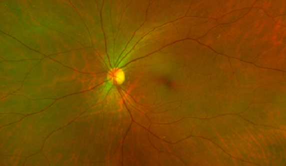
Normal optic nerve
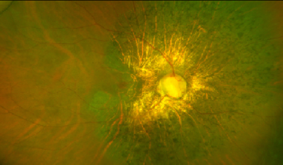
Glaucomatous optic nerve
 As the peripheral visual field narrows, vision declines, and because patients have no symptoms until late stages, diagnosis is often delayed.



FAQ
No. Once the optic nerve is damaged, it does not recover. The goal of treatment is to prevent further optic nerve damage, not to regain lost visual field.
It depends on the tests you receive. If dilation drops are used, additional time is needed for the drops to take effect. We will let you know which tests are planned when you make your appointment.
No. Laser treatment improves aqueous outflow to lower intraocular pressure, and the goal is to prevent further optic nerve damage.
Because the peripheral field narrows first, central vision may feel unchanged for a while. Since there are no noticeable symptoms until the late stage, diagnosis is often delayed, so it is worth checking before symptoms appear.
After dilation, you may be sensitive to light and have difficulty seeing up close for a time. If you plan to drive that day, please let us know when you book.
No. Glaucoma treatment usually starts with medication; laser treatment is done when medication is difficult to use. We cannot say whether it is appropriate without test results.
Like diabetes or hypertension, it requires lifelong management. We check intraocular pressure and visual fields at set intervals to see whether progression has stopped.
We do not judge by pressure alone. Because optic nerve damage can also progress due to impaired blood flow to the nerve, we examine the optic nerve appearance and visual field together.
Not necessarily. Glaucoma requires lifelong care, so even after laser treatment we may continue medication while monitoring how well your pressure is controlled.
We look at intraocular pressure, the optic nerve, and the visual field together. We do not decide based on a single value.
It can be difficult to tell whether the disease is progressing from a single result, so we may repeat the measurement after an interval. We will decide together when to return based on the results.
It varies by person. The course depends on how much the pressure decreases and the extent of prior optic nerve damage, so we do not give a fixed timeframe.
The risk is higher if glaucoma runs in your family. Even without symptoms, we recommend having your intraocular pressure and optic nerve checked.
Before vision correction surgery, we need to check not only the cornea but also the tear film, intraocular pressure, and retina to plan safely.
Glaucoma treatment usually starts with medication, but in some cases, such as congenital glaucoma, surgery is performed first. We decide based on the test results.
When glaucoma is suspected, we first determine whether it is actually present using optical coherence tomography (OCT), fundus photography, and visual field testing. After confirmation, we monitor every 6 months to see if there is progression, because we do not judge based on a single result.
Because glaucoma narrows the peripheral field first. Even when central vision feels fine, people notice peripheral loss late. So we measure separately where and how much you can see, and interpret it together with other findings.
Optic nerve damage is also explained by mechanisms in which blood flow to the optic nerve is impaired, leading to progression. Therefore, we do not judge based on intraocular pressure alone.
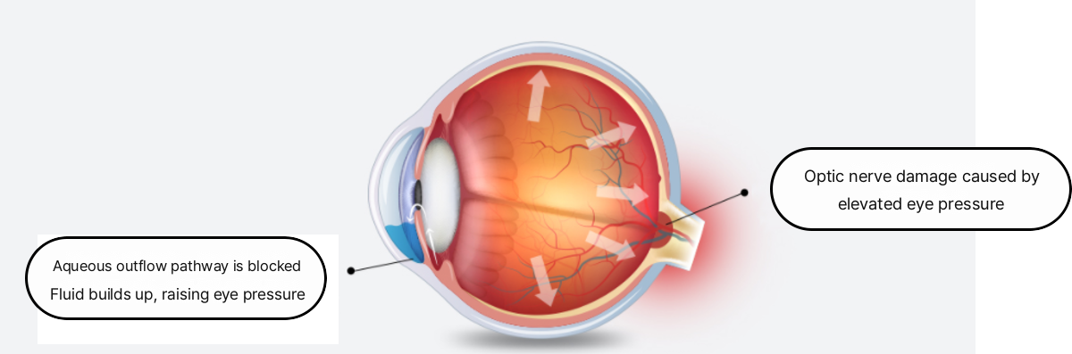
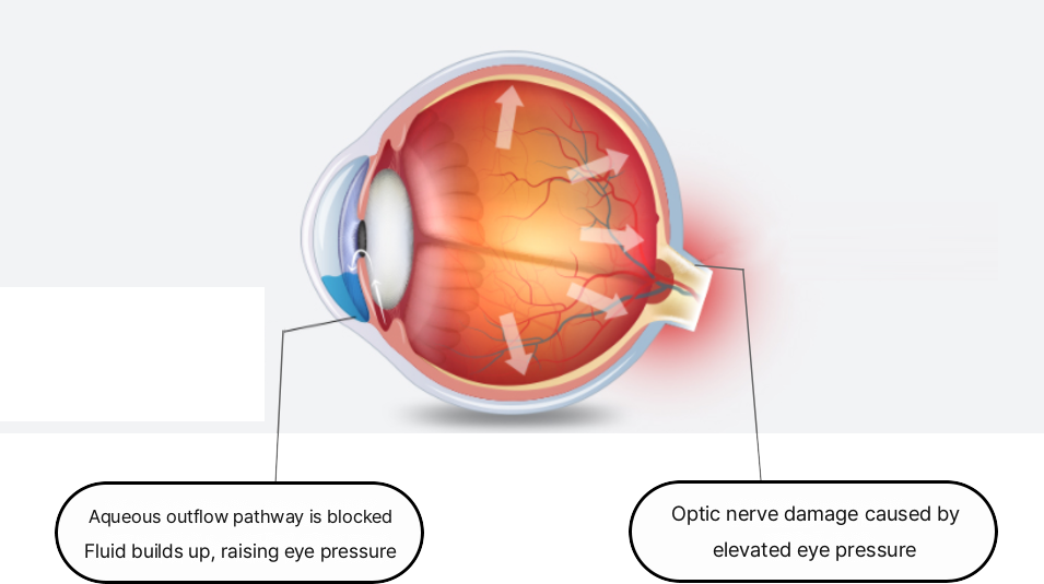
When intraocular pressure rises, the optic nerve can be compressed and damaged, so it is considered a major cause of glaucoma. Lowering it is also a primary goal of treatment. However, we do not make decisions based on pressure alone; we interpret it together with the optic nerve and the visual field.
This finding suggests there may have been damage. We check its extent with a visual field test to see how far it has progressed. Because a damaged optic nerve does not recover, we set a baseline and compare changes over time.


