Directions

KakaoTalk

Booking

YouTube

Instagram

Blog

We diagnose and treat
conditions that can threaten vision
Because retinal care is complex and critical,
it requires specialized clinicians and advanced equipment.
Because retinal diseases can ultimately lead to blindness, early diagnosis and
ongoing treatment and management are important.
At Gangnam Central Eye Clinic’s Retina Clinic, diagnostic equipment and
our ophthalmologists assess each patient’s condition step by step,
and we aim to choose a treatment suited to that condition.

GANGNAM CENTRAL EYE CLINIC
Who Should Have Regular Retinal Exams
Even if you have no particular symptoms, we recommend regular eye examinations from your 40s onward.
01

Patients with diabetes

02

Patients with high blood pressure

03

Patients with high myopia

04

Age 50+
Middle-aged and older adults

05

Those who notice decreased vision
or visual disturbances

06

Those
experiencing changes in their visual field

If you have retinal symptoms
Get an eye examination before it’s too late
If you are in a retinal disease risk group
Detect it early with preventive eye examinations
GANGNAM CENTRAL EYE CLINIC
Gangnam Central Eye Clinic’s Systematic Retina Treatment System

Same-Day Exam · Same-Day Treatment 1Day System

Based on examinations with modern equipment and our doctors’ diagnosis,
we propose a treatment plan suited to each patient.

GANGNAM CENTRAL EYE CLINIC
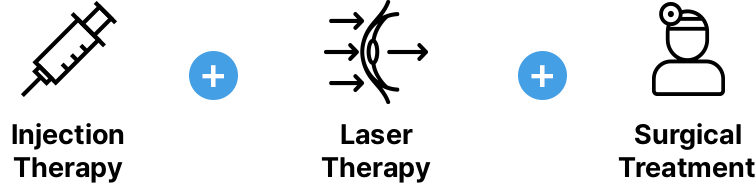
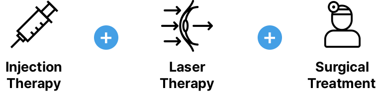
Intravitreal Injection Therapy
Drug therapy is a procedure that improves microvascular circulation in the retina.
Anti-VEGF injections are given directly into the vitreous cavity after the eye is numbed with anesthetic eye drops.
Procedure Steps
After the area around the eye is disinfected,
anesthetic eye drops are applied, and a fine ophthalmic needle is used to
inject the medication into the vitreous cavity.
Direct Intravitreal Drug Delivery
Intravitreal injection has been performed for a long time;
by delivering medication directly into the eye, it reduces the amount used and
minimizes side effects from the drug, making it a safe treatment method.
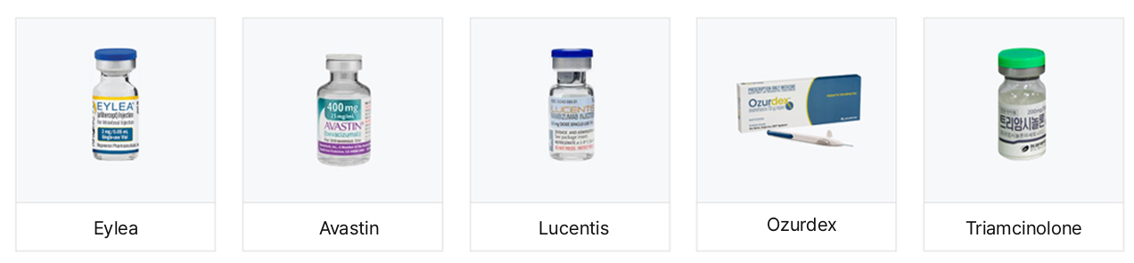
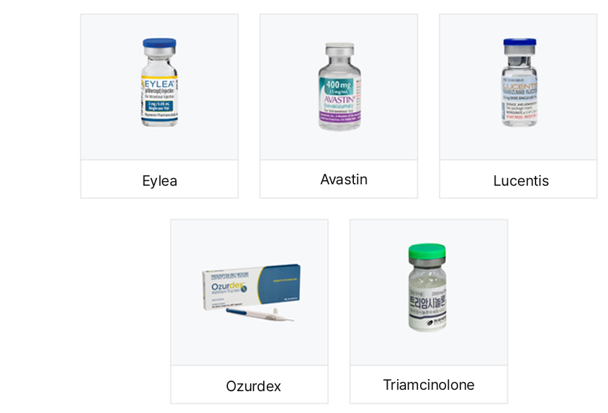
Anti-vascular endothelial growth factor agents such as Lucentis, Avastin, and Eylea, and
steroid agents such as Ozurdex and triamcinolone can be used. The drug is selected for injection according to the pattern of macular degeneration and the degree of edema.

GANGNAM CENTRAL EYE CLINIC
Laser Therapy
Retinal laser therapy applies laser to areas with insufficient retinal blood circulation,
reduces the retina’s oxygen demand, and
is often used to help prevent harmful new blood vessels from forming.
Focal Laser Therapy
Used to reduce swelling in diabetic macular edema,
and to treat serous retinal detachment in central serous chorioretinopathy.
A mild laser is applied to the macula, so there is little pain.
Photodynamic Therapy
For wet macular degeneration and in cases where focal laser therapy is difficult,
it is a specialized laser therapy. There is no pain during the procedure, but
sun exposure is restricted for 5 days.
Panretinal Photocoagulation (PRP)
To suppress neovascularization in proliferative diabetic retinopathy,
the entire retina except the macula is
treated with laser photocoagulation.
Barrier Laser Therapy
A laser treatment performed to prevent progression of retinal tears or localized retinal detachment;
there is pain during the procedure.
GANGNAM CENTRAL EYE CLINIC
Surgical Treatment
Performed when medication or laser therapy is ineffective.
Surgical treatment creates a drainage pathway so that aqueous humor inside the eye can
appropriately exit along the outer wall of the eyeball.
Scleral Buckling
In rhegmatogenous retinal detachment caused by a retinal hole,
scleral buckling may be performed, pressing the eye from the outside to treat the detachment.
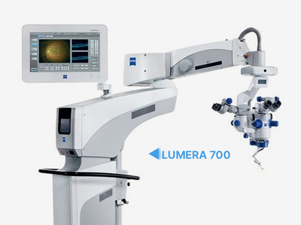
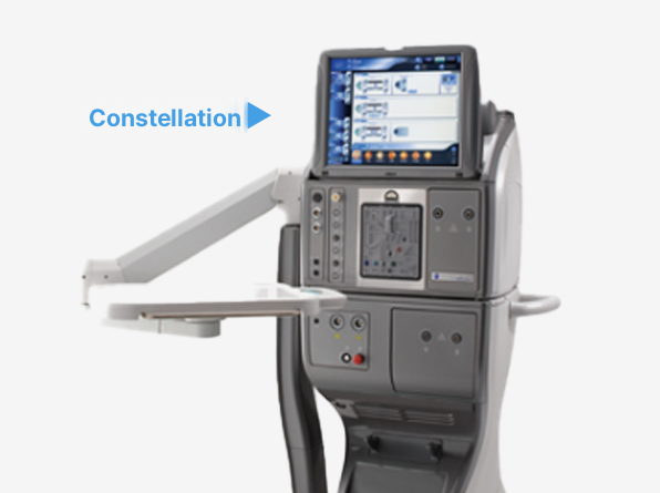
Vitrectomy
For retinal diseases, intravitreal injections or laser treatment are first considered, but if vitreous hemorrhage (bleeding inside the eye)
persists for a considerable period or tractional retinal detachment develops along with changes in the retinal nerve tissue, vitrectomy is performed.
It is a major surgery in ophthalmology; generally, the blood-filled vitreous is removed and aspirated, and
the vitreous cavity is filled with fluid or gas. After surgery, it is important to rest and recover.

FAQ

Retinal Treatment FAQs

Is there anything I should tell you before the exam?

Please let us know about any medications you take and any systemic conditions you have. If dilating eye drops are used, your eyes may be sensitive to light for a while after the exam, so we will also check your plans for that day.

I have diabetes, but my eyes seem fine.

Diabetic retinopathy has no early symptoms, so without regular checks the right time for treatment is often missed. Even without symptoms, you should have a retinal exam once a year.

What tests are done in a retinal checkup?

After dilation, we examine the fundus to check the macula and periphery, and we use optical coherence tomography to view cross-sections of the retina and choroid. If needed, fluorescein angiography identifies new vessels and leakage, and if the inside of the eye is cloudy, ultrasound is used.

If something is found on the exam, how often should I return?

If suspicious findings appear, we confirm with fundus examination and optical coherence tomography, then usually monitor your condition at 6-month intervals. The interval varies by findings, and we will advise you during your visit.

Should I get a retinal exam even if I have no symptoms?

If you have high myopia, diabetes, or high blood pressure, or are over 40, we recommend a retinal exam even without symptoms. Even without particular symptoms, it is good to have regular checkups from your 40s onward.

If I see fly-like specks floating in front of my eyes, should I be examined?

Symptoms such as floaters (seeing dust or gnats), flashing lights, or a portion of the visual field appearing obscured indicate the need for a retinal exam. We check the retina with a fundus exam and optical coherence tomography.

A health checkup showed an abnormal retinal finding.

If you were told you may have a retinal disease such as macular degeneration, epiretinal membrane, or diabetic retinopathy, it is advisable to have a detailed fundus examination and optical coherence tomography. Depending on the findings, we perform regular follow-up every 6 months.

Why should retinal diseases be found early?

The retina is neural tissue, so once damaged it is difficult to restore. That is why early detection and steady management are important.

If my vision has declined or the range I can see seems to have changed, should I get a retinal exam?

If your vision has declined, you have difficulty seeing, or you notice changes in your visual field, we recommend regular retinal exams. We evaluate the retina with a fundus exam and optical coherence tomography.

Do you check the retina after cataract surgery?

Yes. Between 3 and 6 months after cataract surgery, we recheck the retina and look for posterior capsule opacification (secondary cataract). After that, annual checkups screen for coexisting conditions such as glaucoma and macular degeneration.

How are the retinal exam and the glaucoma exam different?

For the retina, we assess its condition with a fundus examination and optical coherence tomography. For glaucoma, we confirm the diagnosis by adding a visual field test to optical coherence tomography and fundus photography.

Online Consultation