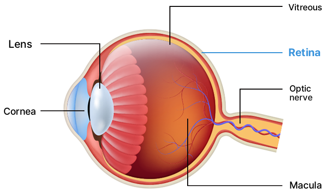

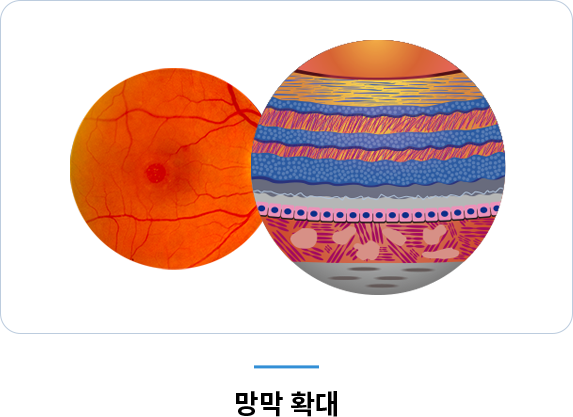
 Retina magnifiedCross-section of the eye
FAQ
Because the retina is neural tissue, damage to it is irreversible. That is why regular eye examinations are important for catching problems early.
Floaters are a symptom, not a disease in themselves. However, they can be a sign of conditions that cause retinal detachment, retinal tears, or retinal hemorrhage; if the symptoms recur, you should have them checked.
Even when diabetic retinopathy has progressed considerably inside the eye, normal vision may be maintained if the macula is unaffected. However, progression does not stop, so you should have a retinal exam once a year even without symptoms.
Depending on the symptoms, we select from fundus examination and photography, optical coherence tomography, fluorescein angiography, ICG angiography, ultrasound, and visual field testing.
Retinal detachment is an emergency. If part of your visual field is increasingly obscured or your vision drops suddenly, do not delay and contact us.
This can occur with an epiretinal membrane, which forms a thin sheet over the macula and tugs on the nerve layers, or with macular degeneration. You can check simply with an Amsler grid; if the grid lines look distorted, we recommend a detailed examination.
This is called photopsia. It occurs when the vitreous separates from the retina and tugs on it, and it may appear together with floaters. Because it can signal a retinal tear or retinal detachment, if the symptoms recur you should have it checked.
It is divided into dry and wet types. Dry refers to early-stage changes such as drusen or retinal pigment epithelium atrophy, with gradual vision loss, while wet involves exudation that distorts the macular structure and most often progresses acutely. There may be no subjective symptoms in the early stage, so it is often found during routine exams.
If macular degeneration, epiretinal membrane, or diabetic retinopathy is suspected, we confirm whether the finding is present with a detailed fundus exam and optical coherence tomography (OCT). Follow-up is then performed every 6 months according to the confirmed findings.
We recommend routine eye examinations after age 40 even if you have no particular symptoms. If you have high myopia, diabetes, or hypertension, or if you notice changes in vision or visual field, we recommend getting checked regardless of age.
To view the fundus, we dilate the pupils with drops, so after the exam your eyes may be sensitive to light and near vision may be blurry for a while. If you plan to drive that day, please let us know when you book.
If you have retinal diseases such as macular degeneration or epiretinal membrane, we do not recommend multifocal intraocular lenses. We first assess the retina and then choose the intraocular lens.
This is sometimes used to describe retinal vascular occlusion, in which the retinal artery or vein becomes blocked. Vision may suddenly decrease or feel clouded like a dark haze, and it is related to hypertension, diabetes, and cardiovascular disease.
This can be a symptom of central serous chorioretinopathy. Exudate from the choroid collects in the macula, causing a coin-shaped shadow or making objects appear smaller and farther away. Recurrence is common, and stress is considered a major contributing factor.
This can occur with a macular hole, in which tissue in the macula thins and a round opening forms. If left untreated it can lead to retinal detachment, so if the symptoms recur we recommend a detailed examination.


