





It shows gland structure, not a complete dry-eye diagnosis. Shortened, distorted, or missing-looking glands may help explain the examination findings, but the clinician still needs to assess oil expression, tear stability, the eye surface, and your symptoms. A normal-looking scan does not rule out poor gland function.
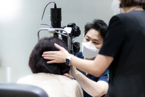
What happens during a meibography test?
The examiner gently turns the eyelid to expose its inner surface and records an infrared image of the glands. Tell the examiner if this is uncomfortable. The image is then assessed for gland shape and areas where normal gland structure is not visible.
Meibography does not measure tear production or the time before tears break up after a blink. Those questions require other measurements. The dry-eye test comparison explains how TBUT, Schirmer testing, staining, and gland expression complement the scan.
What do the meibomian glands do?
Meibomian glands run vertically through the upper and lower eyelids. Their oily secretion forms the outer layer of the tear film and slows evaporation. If openings are blocked or the secretion becomes thick, tears may break up quickly even when tear volume is not extremely low.
Typical complaints include burning, a gritty feeling, fluctuating blur that clears after blinking, contact-lens intolerance, or symptoms that worsen with prolonged screen use. Those symptoms can also have other causes, which is why testing is combined rather than interpreted in isolation.
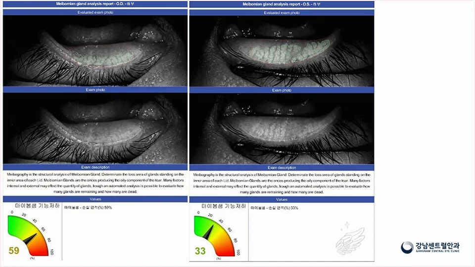
What do gland dropout and other image findings mean?
An infrared image can help the clinician assess gland length, contour, tortuosity, and areas of apparent dropout. It is also useful as a baseline when symptoms and other signs are followed over time.
A percentage or color grade can make the report easy to read, but the number should not be treated as a treatment order. Image quality, eyelid eversion, age, contact-lens history, and the grading method can affect interpretation.
| Finding | What it may indicate | What must still be checked |
|---|---|---|
| Shortened or missing gland area | Structural gland loss | Symptoms, secretion, tear stability, ocular surface |
| Tortuous glands | Altered gland shape | Lid inflammation, obstruction, clinical relevance |
| Dilated ducts | Possible outflow obstruction | Opening appearance and expressibility |
| Asymmetry | Different structure between lids or eyes | Imaging technique and repeatability |
| Normal-looking structure | No obvious structural loss | Function can still be abnormal |
Which tests check function rather than gland structure?
The National Eye Institute describes slit-lamp evaluation, tear-production testing, and tear break-up time as common parts of dry-eye testing. Clinicians may also evaluate staining, lid margins, blink pattern, tear meniscus, and meibomian secretion.
TFOS describes dry eye as a multifactorial disease involving tear-film homeostasis. That is why a single machine result should be connected to the patient's symptoms and the part of the tear system that is not functioning well.
| Test | Main question | Example interpretation |
|---|---|---|
| Tear break-up time | How long does the tear film remain stable after a blink? | Short stability may fit evaporative or mixed dry eye |
| Surface staining | Is the cornea or conjunctiva damaged? | Location and severity influence treatment priority |
| Schirmer or tear volume | Is aqueous tear production reduced? | Helps identify aqueous-deficient components |
| Lid-margin examination | Are openings blocked or inflamed? | Connects the scan to visible lid findings |
| Gland expression | Does oil come out and what is its quality? | Adds functional information to structural imaging |
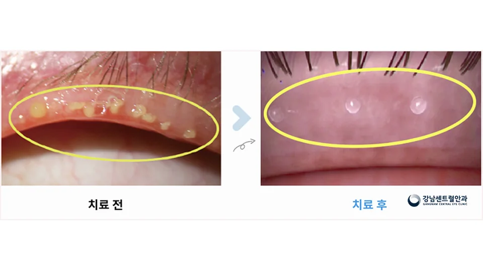
Who may benefit from a broader dry-eye evaluation?
Consider an eye examination when discomfort repeatedly limits reading, screen work, contact-lens wear, or sleep; when vision fluctuates; or when over-the-counter drops provide only brief relief. Tell the clinician which eye is worse, when symptoms peak, and what products you have tried.
Dry eye can be influenced by contact lenses, eyelid inflammation, incomplete blinking, medicines, autoimmune disease, environment, and previous eye surgery. A useful visit looks for the pattern rather than assuming every symptom is caused by blocked glands.
Bring a list of eye drops and medications, your contact-lens schedule, and a short note on when symptoms are worst. This often makes the scan easier to interpret in context.
Does a gland-loss score decide which treatment I need?
No. A gland-loss score describes an image; it does not choose a treatment by itself. Ask the clinician to explain how the score relates to your symptoms and the functional findings rather than treating a percentage as a diagnosis.
Ask whether the main finding is structural loss, poor oil expression, unstable tears, surface damage, or a combination. Also ask which measurement can be repeated to judge whether symptoms and function are improving.
The goal is not to make the image look perfect. The goal is to reduce symptoms, protect the ocular surface, and improve tear-film stability with a plan matched to the findings.
Frequently asked questions
Does gland dropout on meibography mean the glands are permanently gone?
The image may show areas where normal gland structure is not visible. The clinical meaning depends on image quality, grading, symptoms, secretion, and other dry-eye findings; discuss the result with an eye-care professional.
Can meibography diagnose dry eye by itself?
No. It provides structural information. Symptoms, tear stability, tear volume, surface staining, lid findings, and gland expression may all be relevant.
Is the test painful?
Imaging is generally noninvasive, although the eyelid is gently turned to expose its inner surface. Tell the examiner if you are uncomfortable.
Can a normal scan rule out meibomian-gland dysfunction?
No. Glands can look relatively preserved while secretion quality or expressibility is abnormal, so function still needs examination.
Should the scan be repeated?
Repeat imaging may be useful when the clinician needs a comparable baseline, but symptom and functional measures are also important for follow-up.
Medical references
This article was checked against public guidance from medical authorities and professional organizations.
Medical information standards
This article was prepared from publicly available ophthalmology guidance and the clinic's examination process. A diagnosis or treatment plan can only be made after an individual eye examination and consultation.
- Clinic director: Pung GyeHyeon, MD · Ophthalmologist
- Organization: Gangnam Central Eye Clinic · Seoul, Korea
- Published / last updated: 2026-09-13 / 2026-10-03
This article is general medical information and is not a substitute for personal diagnosis or treatment. Seek prompt eye care for sudden vision loss, severe pain, marked redness, flashes, or a new curtain-like shadow.