





No. The National Eye Institute notes that high pressure increases glaucoma risk, yet glaucoma can occur with pressure in the usual range. A useful evaluation interprets the reading with corneal thickness, optic-nerve appearance, OCT, visual-field results, angle anatomy, and previous measurements.
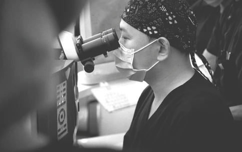
How is eye pressure measured?
Tonometry estimates intraocular pressure. Noncontact instruments use an air pulse and are common in screening. Applanation instruments gently flatten a small area of the anesthetized cornea and are often used for clinical confirmation. Other handheld or rebound devices may be selected for particular situations.
Readings can vary with time of day, squeezing, breath holding, body position, technique, and the biomechanical properties of the cornea. A surprising result may be repeated rather than treated as a permanent label.
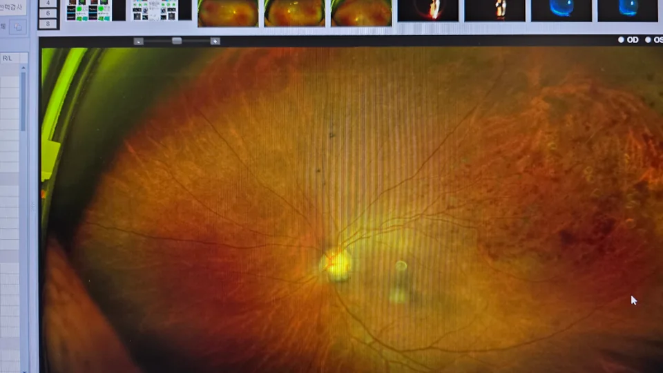
What is considered normal eye pressure?
A commonly cited population range is not a personal safety threshold. The NEI explains that pressure above 21 mmHg is higher than normal for most people, but the amount an individual's optic nerve can tolerate differs.
A person can have ocular hypertension without detected nerve damage, or normal-tension glaucoma with pressure readings in the usual range. Diagnosis and treatment decisions therefore do not rest on one cutoff.
Ask what the number means for your optic nerve, corneal thickness, family history, age, and previous readings.
Which tests complete a glaucoma evaluation?
Glaucoma is a group of diseases that damage the optic nerve. Clinicians compare structural tests with functional testing and use repeated examinations to decide whether change is occurring.
A colored OCT map is not a diagnosis by itself. High myopia, disc size, segmentation error, and image quality can affect the report, just as attention and learning effects can affect a visual-field test.
| Test | What it contributes | Why trends matter |
|---|---|---|
| Tonometry | Pressure at the time of measurement | Pressure varies and risk is individual |
| Pachymetry | Central corneal thickness | Supports interpretation of pressure and risk |
| Optic-nerve examination or photography | Disc appearance and baseline documentation | Allows comparison over time |
| OCT | Retinal nerve-fiber and ganglion-cell structure | Repeatable loss can support progression assessment |
| Visual field | Functional sensitivity across central and peripheral vision | Confirms whether function is changing |
| Gonioscopy | Drainage-angle anatomy | Helps distinguish open and narrow or closed angles |
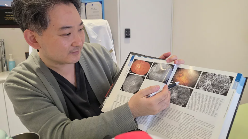
Who should discuss a full glaucoma examination?
Risk increases with factors such as family history, age, higher eye pressure, suspicious optic-nerve appearance, high myopia, certain ancestry, steroid exposure, and some medical conditions. The recommended interval is individualized.
Open-angle glaucoma is often symptomless early. Waiting for tunnel vision or reduced central acuity can delay detection, so risk-based examination is more useful than symptom-based reassurance.
- A parent or sibling with glaucoma
- Previous high-pressure or suspicious optic-nerve result
- High myopia or prior eye surgery
- Use of steroid eye drops, inhalers, tablets, injections, or skin preparations
- Diabetes, vascular conditions, or a clinician's recommendation for monitoring
Which symptoms require urgent care?
Most chronic open-angle glaucoma does not cause an acute painful episode. Sudden severe eye pain, headache, nausea, halos, marked redness, and rapid vision change can indicate an acute angle-closure event or another emergency.
Do not wait for a routine appointment when symptoms are sudden or severe. Seek urgent local eye care.
Sudden severe eye pain, headache with nausea, halos and a red eye, or rapid vision loss require urgent assessment.
Frequently asked questions
What is a normal eye pressure?
A population range can be a reference, but there is no single safe number for every optic nerve. Your reading must be interpreted with corneal thickness, optic-nerve findings, visual field, and risk factors.
Can I have glaucoma with normal eye pressure?
Yes. Normal-tension glaucoma can damage the optic nerve even when measured pressure is within the usual range.
Does high eye pressure mean I have glaucoma?
Not necessarily. Some people have ocular hypertension without detected optic-nerve damage, but they may need risk assessment and monitoring.
Does the air-puff test hurt?
It is noncontact and brief, though the puff can be startling. A clinician may confirm the result with another tonometry method.
How often should eye pressure be checked?
The interval depends on age, family history, previous readings, optic-nerve findings, medications, and other risks. An eye-care professional should set the schedule.
Medical references
This article was checked against public guidance from medical authorities and professional organizations.
Medical information standards
This article was prepared from publicly available ophthalmology guidance and the clinic's examination process. A diagnosis or treatment plan can only be made after an individual eye examination and consultation.
- Clinic director: Pung GyeHyeon, MD · Ophthalmologist
- Organization: Gangnam Central Eye Clinic · Seoul, Korea
- Published / last updated: 2026-09-13 / 2026-09-13
This article is general medical information and is not a substitute for personal diagnosis or treatment. Seek prompt eye care for sudden vision loss, severe pain, marked redness, flashes, or a new curtain-like shadow.